About Me

I’m a Ph.D. in Biomedical Engineering (University of Ottawa, 2024) with a passion for translating complex scientific concepts into clear, impactful narratives. With a B.Sc. in Nanoscience and M.Sc. in Biophysics from the University of Guelph, I’ve built over a decade-long career in biomedical research, specializing in biomaterials, advanced imaging (AFM, SEM, Raman, optical microscopy), and nanoparticle therapeutics. My writing reflects this expertise, spanning peer-reviewed publications, technical reports, and accessible science communication for diverse audiences.

My research journey includes pioneering biomaterials for orthopedic implants to enhance osseointegration in diabetic patients, resulting in 6 peer-reviewed publications, and exploring wound healing innovations to combat biofilm formation. As a Nanoparticle Chemistry Technician at the University of Calgary, I contributed to cutting-edge nanoparticle therapies for autoimmune diseases. Beyond academia, I’m an entrepreneur and content creator through Mint 'n Scrypt, where I write about Bitcoin, digital assets, markets, and the intersection of science and technology.

On this portfolio, you’ll find my work on scientific articles and conference presentations to engaging pieces on healthcare innovation and emerging technologies. Based in Calgary, Alberta, I’m driven to bridge science and storytelling, whether through medical writing, science communication, or thought leadership in life sciences and beyond. Explore my portfolio to see how I bring clarity and impact to complex ideas.

Journal Articles

Exploring the influence of anodization-derived nanotubular and honeycomb surfaces on the osteogenic behaviour of human MG63 osteoblastic cells

Nanoscale topography plays a pivotal role in modulating cellular behavior and has been a key parameter in
the design of cell-instructive surfaces for biomedical applications. This study investigates the differential
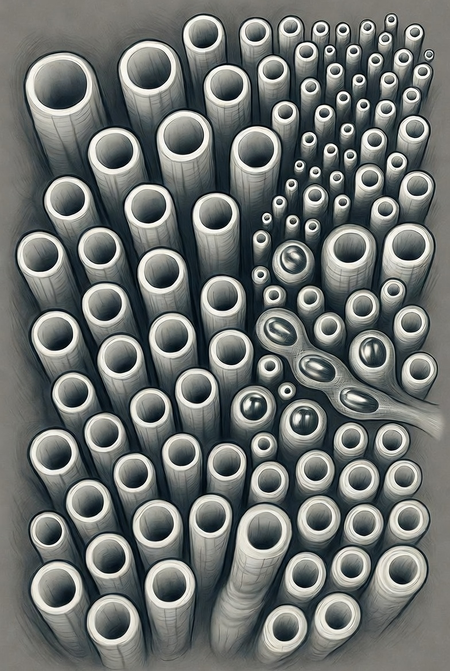
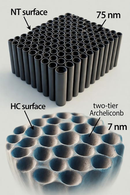
effects of two anodized titanium surfaces – a conventional nanotubular (NT) surface (∼75 nm diameter) and a two-tier honeycomb (HC) architecture – on the response of human MG63 osteoblastic cells. The HC surface, characterized by higher spatial entropy and a complex arrangement of smaller nanotubes (∼7 nm in diameter) clustered within larger domains (∼109 nm in diameter), significantly enhances early cellular
functions, including proliferation, viability and upregulation of osteogenic markers (RUNX2, OSX, ALP), with the YAP/Hippo pathway likely implicated as a key mediator. This is evidenced by increased focal adhesions and nuclear YAP1 localization, underscoring the HC surface's capacity to promote cellular attachment and early differentiation. Conversely, the NT surface, with its more ordered nanotube array, induces comparable mineralization but yields higher-quality mineral deposits enriched with crystalline hydroxyapatite, suggesting greater efficacy in supporting mature mineral formation. These findings highlight the selective influence of nanotopographical features on early cellular dynamics versus long-term mineralization, offering critical insights into structure–function relationships governing MG63 cellular response to anodized titanium. By demonstrating the HC surface's prowess in early osteogenesis and the NT surface's strength in stable mineral deposition, this research advances the design of cell-instructive biomaterials tailored to distinct phases of bone regeneration, with implications for tissue engineering and biomedical implant technology.

Nanotubular Gradients on Titanium: High-Throughput Screening of Nanoscale Architectures of Variable Topographical Complexity

Advancements in cell-instructive biomaterials hinge on the precise design of their nanoscale topography, a critical factor in controlling cell–surface interactions. Nanofabrication techniques such as e-beam and nanoimprint lithography enable accurate nanopatterning on a wide range of materials. However, their limited applicability and scalability to medically relevant metals such as titanium, hinder the creation and modulation of precisely designed nanotopographies on metallic substrates to inve...

Investigating the interplay between environmental conditioning and nanotopographical cueing on the response of human MG63 osteoblastic cells to titanium nanotubes

R. Berthelot and F. Variola,
Biomater. Sci., 2025, 13, 946

DOI: 10.1039/D4BM00792A


To request permission to reproduce material from this article, please go to the
Copyright Clearance Center request page.


If you are an author contributing to an RSC publication, you do not need to request permission
provided correct acknowledgement is given.


If you are the author of this article, you do not need to request permission to...

Nanoscale imaging approaches to quantifying the electrical properties of pathogenic bacteria

Biofilms are natural, resilient films formed when microorganisms adhere to a surface and form a complex three-dimensional structure that allows them to persist in a wide variety of environments. Readily forming in hospitals and on medical equipment, biofilms are frequent causes of infections and their subsequent complications. Due to the complexity of these structures, systematically studying individual bacterial cells and their interactions with their surrounding environment will provide a deeper understanding of the processes occurring within the biofilm as whole versus bulk population based methods that do not differentiate individual cells or species. Methods based on atomic force microscopy (AFM) are particularly suited to the study of individual cells, but are underutilized for the study of bacterial electrical properties. The ability of electrical currents to impair bacterial attachment is well documented, but to utilize electrical current as an effective antibacterial treatment, it is important to understand the electrical properties of bacteria. Therefore, we used AFM, Kelvin probe force microscopy, and ResiScope (module to perform conductive AFM) to measure the surface potential and conductance of Psuedomonas aeruginosa and methicillin resistance Staphylococcus aureus (MRSA) on gold and stainless steel. This is the first study to directly measure the electrical resistance of single bacterial cells using ResiScope. Our goal was to develop a framework for measuring biological molecules using conductive AFM. We found that the average resistance for P. aeruginosa was 135 ± 25 GΩ, while MRSA had an average of 173 ± 16 GΩ. Using KPFM, the surface potential of MRSA shifted from −0.304 V to 0.153 V and from −0.280 V to 0.172 V for P. aeruginosa on gold versus stainless steel substrates, respectively. In an attempt to identify a potential charge carrier, peptidoglycan was also measured with the ResiScope module and shown to have a resistance of 105 GΩ.

Electroceutical Approach for Impairing the Motility of Pathogenic Bacterium Using a Microfluidic Platform

All articles published by MDPI are made immediately available worldwide under an open access license. No special
permission is required to reuse all or part of the article published by MDPI, including figures and tables. For
articles published under an open access Creative Common CC BY license, any part of the article may be reused without
permission provided that the original article is clearly cited. For more information, please refer to
https://www.mdpi.com/...

Harnessing electrical energy for anti-biofilm therapies: effects of current on cell morphology and motility

Preferentially altering bacterial migration could be a successful approach for augmenting the natural wound-healing process. Inducing electrotactic behaviours through the application of physiologically safe currents is one possible strategy for altering bacterial movement. By controlling bacterial movement at the site of infection, healing times and the severity/extent of bacterial infection could be reduced. Here, we deployed microfluidics and atomic force microscopy to determine the ef...